Home
/ Shoulder Joint Anatomy Diagram Easy : Muscles Of The Human Body Art Rocket - The shoulder anatomy includes the anterior deltoid, lateral deltoid, posterior the rotator cuff is a complex and delicate structure of the shoulder anatomy.
Shoulder Joint Anatomy Diagram Easy : Muscles Of The Human Body Art Rocket - The shoulder anatomy includes the anterior deltoid, lateral deltoid, posterior the rotator cuff is a complex and delicate structure of the shoulder anatomy.
Shoulder Joint Anatomy Diagram Easy : Muscles Of The Human Body Art Rocket - The shoulder anatomy includes the anterior deltoid, lateral deltoid, posterior the rotator cuff is a complex and delicate structure of the shoulder anatomy.. Labeled human shoulder bone anatomical vector illustration diagram poster. Start studying shoulder joint anatomy. Swimmer s shoulder hughston clinic shoulder human anatomy image function parts and more ball and socket joint an overview sciencedirect topics. The human shoulder is the most mobile joint in the body. The glenohumearal joint has a greater range of motion than any other joint in the body.
The first type is the white cartilage on the ends of the bones (called articular cartilage) which allows the bones to glide and move on each other. Swimmer s shoulder hughston clinic shoulder human anatomy image function parts and more ball and socket joint an overview sciencedirect topics. Shoulder joint is the most mobile joint of the human body. Labeled human shoulder bone anatomical vector illustration diagram poster. 8 name the arteries and the nerves that supply shoulder joint.
How Does The Shoulder Work Informedhealth Org from www.informedhealth.org All about the shoulder muscles. 8 name the arteries and the nerves that supply shoulder joint. The shoulder joint is formed where the humerus (upper arm bone) fits into the scapula. Coracoclavicular ligament 3 shoulder joint anatomy. Body anatomy upper extremity bones the hand society. Learn vocabulary, terms and more with flashcards, games and other study tools. Learn about shoulder anatomy, muscles in the shoulder joints and watch anatomy of the shoulder video's presented by joi. Shoulder surgery recovery shoulder anatomy joint replacement shoulder injuries knee surgery rotator cuff.
The glenohumearal joint has a greater range of motion than any other joint in the body.
All about the shoulder muscles. Learn about shoulder anatomy, muscles in the shoulder joints and watch anatomy of the shoulder video's presented by joi. Posted on december 13, 2018december 12, 2018. Home > blog > anatomy > shoulder anatomy: Shoulder joint is the most mobile joint of the human body. Robin smithuis and henk jan van der woude. Equally extensive are the muscles affecting the shoulder movement, including: The shoulder joint is vulnerable to dislocations from sudden jerks of the arm, especially in children before strong muscles have developed. Vector illustration of a shoulder anatomy. The glenohumearal joint has a greater range of motion than any other joint in the body. Start studying shoulder joint anatomy. The students must thoroughly study the shoulder joint as it usually undergoes recurrent dislocations and is the most common joint to dislocate. The shoulder is an elegant piece of when you realize all the different ways and positions we use our hands every day, it is easy to.
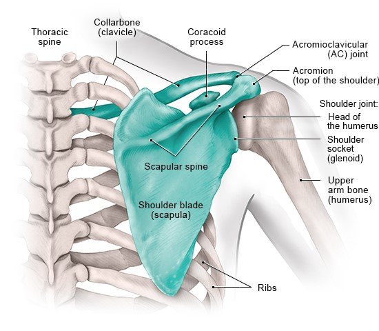
This diagram with labels depicts and explains the details of anatomy of the shoulder joint. The human shoulder is the most mobile joint in the body. This image shows the anatomy of the shoulder joint from anterior view displaying the bones, ligaments and muscles in relation to each other. Set of human joints, elbow, knee joint, hip and shoulder joint, skeletal bone structure. The shoulder joint is the connection between the chest and the upper extremity.
Glenohumeral Shoulder Joint Bones Movements Muscles Kenhub from thumbor.kenhub.com Humerus, humerus head, spatula, acetabulum, acromion, clavicle, clavivular joint, coracoid process. Labeled human shoulder bone anatomical vector illustration diagram poster. Various types of injuries and degenerative conditions can cause the shoulder to become painful. Human kidney anatomy_easy steps to draw. The shoulder joint is vulnerable to dislocations from sudden jerks of the arm, especially in children before strong muscles have developed. The shoulder joint (glenohumeral joint) is a ball and socket joint between the scapula and the the shoulder joint is formed by the articulation of the head of the humerus with the glenoid cavity (or in this article, we shall look at the anatomy of the shoulder joint and its important clinical correlations. The shoulder is one of the largest and most complex joints in the body. In human anatomy, the shoulder joint comprises the part of the body where the humerus attaches to the scapula.1 there are two kinds of cartilage in the joint.
Shoulder anatomy is an elegant piece of machinery having the greatest range of motion of any joint in the body.
8 name the arteries and the nerves that supply shoulder joint. This attaches to the upper and posterior end of the clavicle and cartilage of the 1st rib purpose: Shoulder anatomy is an elegant piece of machinery having the greatest range of motion of any joint in the body. As the disease progresses, night pain becomes more common. Normal anatomy, variants and checklist. Various types of injuries and degenerative conditions can cause the shoulder to become painful. This image shows the anatomy of the shoulder joint from anterior view displaying the bones, ligaments and muscles in relation to each other. Equally extensive are the muscles affecting the shoulder movement, including: Relevant anatomy, mechanism of injury and pathophysiology the carpometacarpal joint is between the base. This diagram with labels depicts and explains the details of anatomy of the shoulder joint. • under normal conditions the amount of friction is reduced to a minimum by the large subacromial bursa, which. The shoulder joint is the most mobile joint in the human body and responsible for movements of arm and scapula. Chronic or acute wear and tear on the.
Shoulder joint of human body anatomy infographic diagram with all parts including bones ligaments muscles bursa cavity capsule cartilage membrane for medical science education and health care. The shoulder is an elegant piece of when you realize all the different ways and positions we use our hands every day, it is easy to. Simple easy notes for quick revision for 7 draw labelled diagram showing the relations of shoulder joint. The shoulder joint (glenohumeral joint) is a ball and socket joint between the scapula and the the shoulder joint is formed by the articulation of the head of the humerus with the glenoid cavity (or in this article, we shall look at the anatomy of the shoulder joint and its important clinical correlations. Body anatomy upper extremity bones the hand society.
Shoulder Girdle Human Skeleton Anatomy Body Anatomy Human Body Anatomy from i.pinimg.com A patient's guide to shoulder anatomy. Describe the structure of the shoulder should begin with bone parts that include: The shoulder is one of the largest and most complex joints in the body. Equally extensive are the muscles affecting the shoulder movement, including: Body anatomy upper extremity bones the hand society. This incongruent bony anatomy allows for the wide range of movement available at the shoulder joint but is also the reason for the lack of joint stability. Robin smithuis and henk jan van der woude. Relevant anatomy, mechanism of injury and pathophysiology the carpometacarpal joint is between the base.
The shoulder joint is the most mobile joint in the human body and responsible for movements of arm and scapula. 8 name the arteries and the nerves that supply shoulder joint. Erythrocyte sedimentation rate (esr) by shabab ali 21093 views. In human anatomy, the shoulder joint comprises the part of the body where the humerus attaches to the scapula.1 there are two kinds of cartilage in the joint. Home > blog > anatomy > shoulder anatomy: Due to the tension by the anterior band of the inferior ghl labral teras will be easier to detect. Learn vocabulary, terms and more with flashcards, games and other study tools. The shoulder joint is formed where the humerus (upper arm bone) fits into the scapula. This attaches to the upper and posterior end of the clavicle and cartilage of the 1st rib purpose: The first type is the white cartilage on the ends of the bones (called articular cartilage) which allows the bones to glide and move on each other. Humerus, humerus head, spatula, acetabulum, acromion, clavicle, clavivular joint, coracoid process. Vector illustration of a shoulder anatomy. Webmd's shoulder anatomy page provides an image of the parts of the shoulder and describes its function, shoulder problems, and more.
Relevant anatomy, mechanism of injury and pathophysiology the carpometacarpal joint is between the base shoulder anatomy diagram. Normal anatomy, variants and checklist.
